We use advanced diagnostic and treatment technologies to evaluate your eye health in greater detail from the retina and optic nerve to the cornea, tear film, and visual field. These objective insights help us detect concerns, understand your unique needs, and tailor your care with greater precision.
Modern eye care requires the ability to identify subtle changes in the eye that may not be visible during a traditional examination alone.
Our practice incorporates advanced diagnostic and therapeutic technologies that allow us to obtain detailed information about the retina, optic nerve, cornea, tear film, visual field, eyelids, and ocular surface.
These technologies help us detect disease earlier, quantify abnormalities, monitor changes over time, and create more individualized treatment plans based on objective measurements rather than symptoms alone.


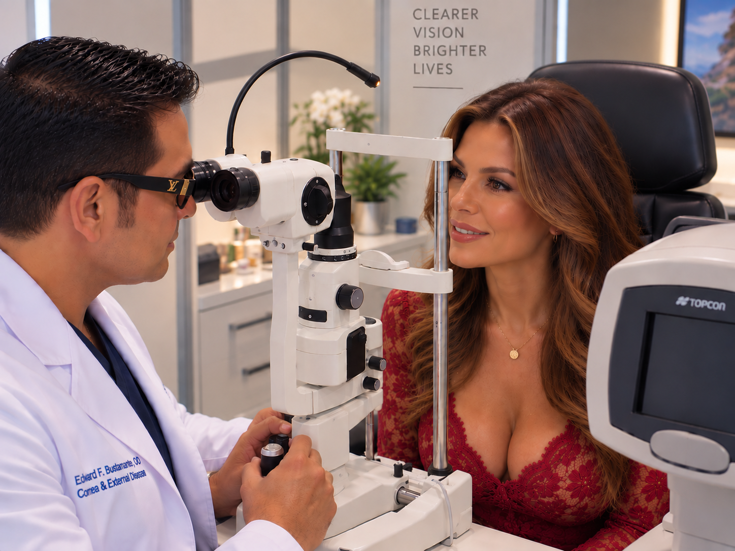
The Topcon Maestro OCT provides highly detailed cross-sectional imaging of structures inside the eye using Optical Coherence Tomography.
It allows us to evaluate the:
Retina
Macula
Retinal nerve fiber layer
Optic nerve
Ganglion cell complex
This technology is especially useful in the diagnosis and monitoring of conditions such as glaucoma, macular degeneration, diabetic retinal disease, and other retinal or optic-nerve abnormalities.
One of the key advantages of OCT is the ability to compare structural measurements over time, helping identify subtle progression even before a patient notices a change in vision.
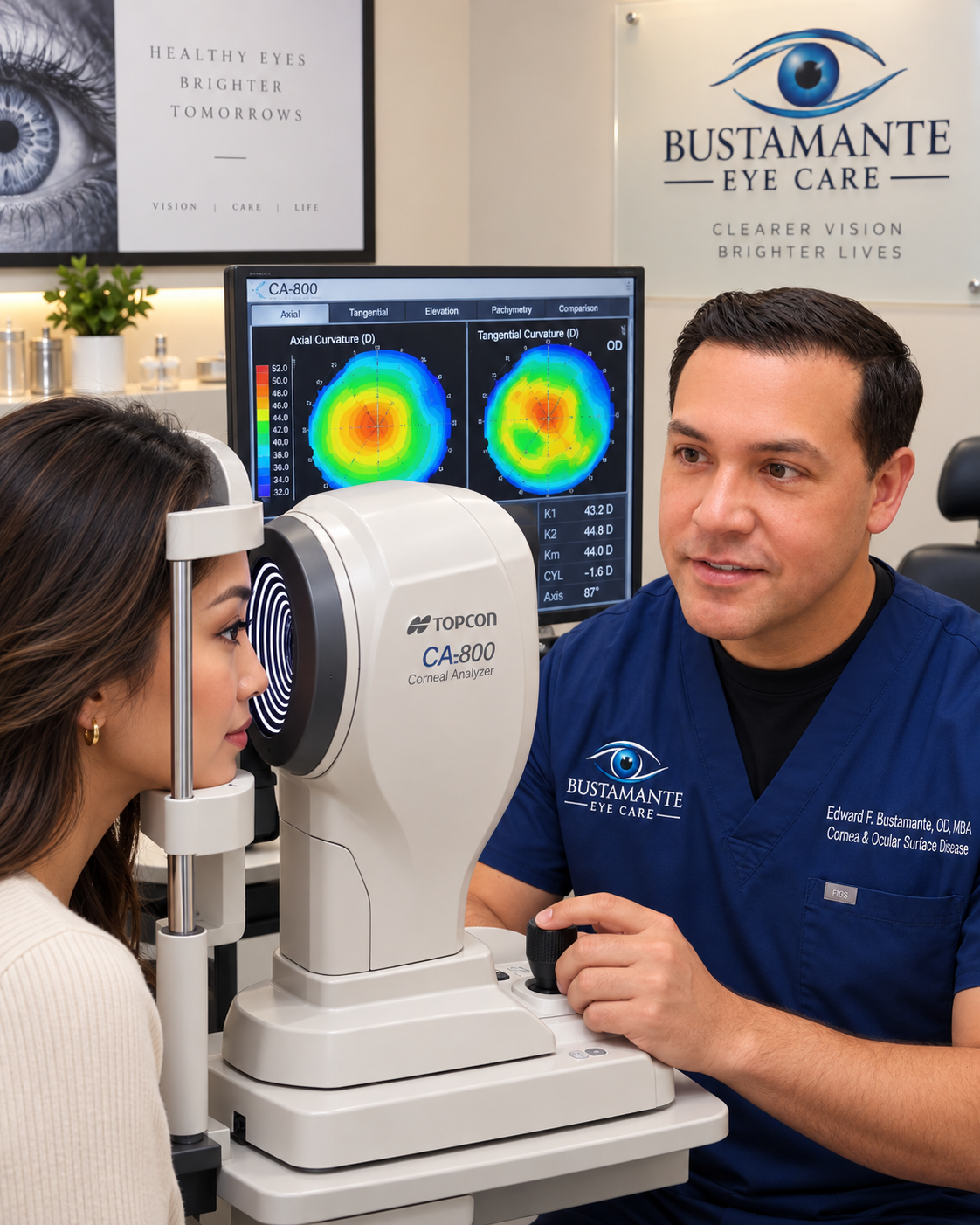
The CA-800 Corneal Topographer provides sophisticated analysis of the shape and optical characteristics of the cornea while also giving us important information about the tear film and ocular surface.

Detailed mapping of corneal curvature and shape.

Identifies corneal surface irregularities.

Measures the tear reservoir along the lower eyelid.

Evaluates tear-film stability after blinking.
The ScoutPro Tear Osmolarity System provides an objective measurement of tear osmolarity.
When the tear film becomes excessively concentrated, or hyperosmolar, it may contribute to ocular-surface stress, inflammation, epithelial damage, and dry-eye symptoms.
Osmolarity testing provides another objective measurement that can be considered together with:
Symptoms
Corneal staining
Tear-film stability
Tear volume
Eyelid and meibomian gland findings
Other ocular-surface measurements
The Humphrey Matrix Visual Field Analyzer measures portions of a patient’s vision that cannot be assessed simply by reading letters on an eye chart.
A patient may have excellent central visual acuity while still developing abnormalities in the peripheral visual field.
Visual-field testing is especially important in evaluating and monitoring glaucoma and other disorders affecting the optic nerve and visual pathway.
What the eye looks like
How vision is functioning
Advanced technology doesn’t just help us identify what’s happening it helps guide how we care for it. This creates a clear separation between diagnostic equipment and treatment technologies.
The eyelids and eyelashes play an important role in maintaining a healthy ocular surface.
Oil, cellular debris, bacterial biofilm, crusting, and inflammatory material can accumulate along the eyelid margins and around the eyelashes. This buildup may contribute to:
Blepharitis
Meibomian gland dysfunction
Eyelid inflammation
Ocular-surface irritation
Evaporative dry eye
Recurrent debris and crusting around the eyelashes
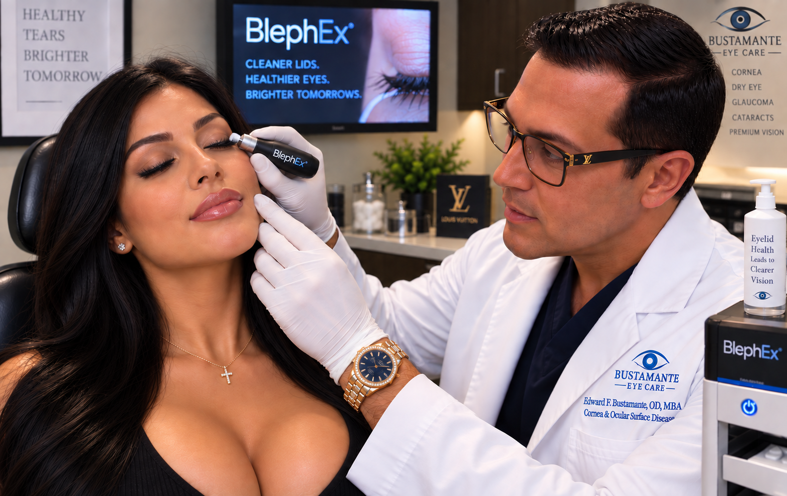
BlephEx is an in-office eyelid cleansing and micro-exfoliation treatment designed to remove accumulated debris and biofilm from the eyelid margins and the base of the eyelashes.
Using a specialized rotating microsponge, the eyelid margins are carefully cleansed to establish a healthier eyelid environment.
The goal is to improve the health of the eyelid margin so other dry-eye and ocular-surface treatments can work more effectively.
“Successful dry-eye treatment often begins with treating the eyelids, because the health of the eyelids and meibomian glands directly influences the quality and stability of the tear film.”
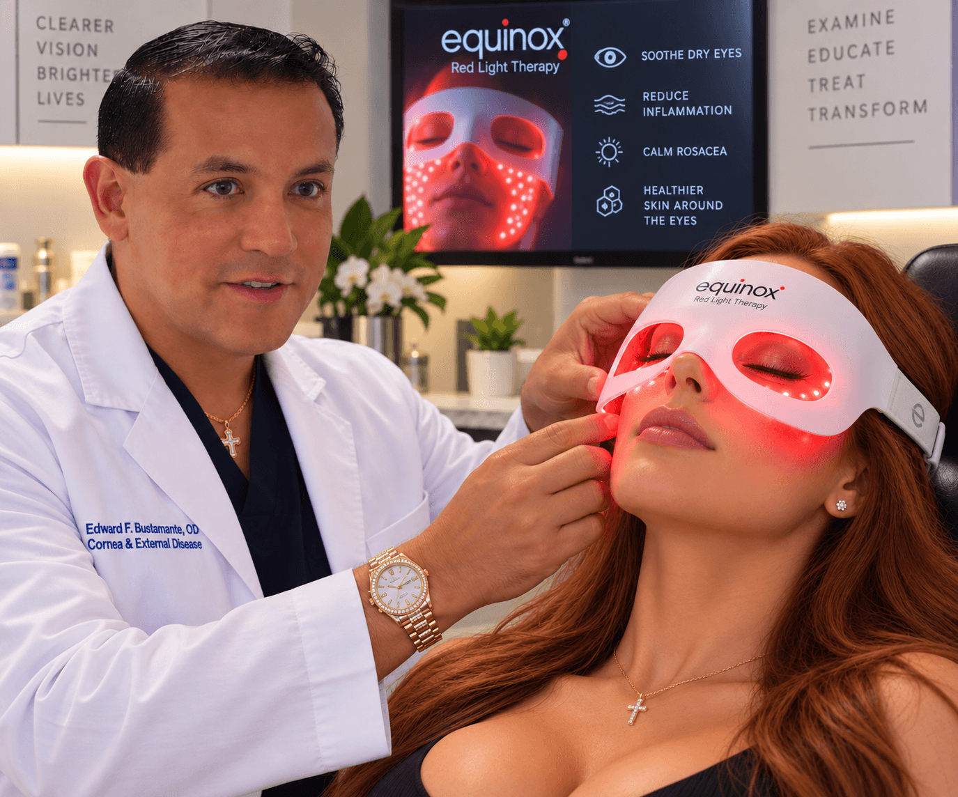
Red-light therapy may be incorporated into treatment programs designed to support eyelid and meibomian-gland function and address inflammatory components of ocular-surface disease.
The goal is to help restore a healthier environment for the tear film and ocular surface rather than simply treating dryness with artificial tears.
Blue-light therapy may be used as part of selected treatment protocols directed toward certain eyelid and microbial-related conditions.
Please keep this description appropriately patient-friendly and avoid making it sound like it is indicated for every dry-eye patient.
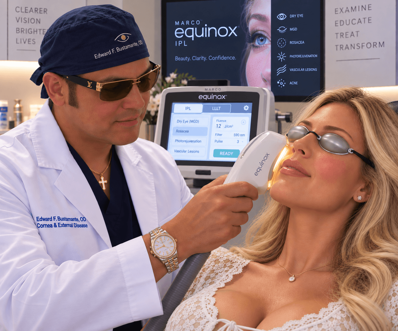
IPL can be incorporated into a comprehensive treatment plan for selected patients with conditions involving:
Meibomian gland dysfunction
Eyelid inflammation
Ocular rosacea
Evaporative dry eye disease
The goal is to address some of the underlying inflammatory and gland-related mechanisms contributing to tear-film instability, rather than simply adding moisture to the eye.
Advanced technology is valuable only when it improves how we understand and care for the patient By combining structural imaging, visual-field testing, corneal mapping, tear-film analysis, osmolarity testing, eyelid cleansing, and advanced ocular-surface treatments, we are able to evaluate the eye from several different perspectives.

What It Helps Evaluate:
Retina & optic-nerve structure

What It Helps Evaluate:
Visual function & peripheral vision

What It Helps Evaluate:
Cornea & tear-film characteristics

What It Helps Evaluate:
Tear-film concentration

What It Helps Evaluate:
Eyelid margin & ocular-surface health

What It Helps Evaluate:
Selected eyelid & ocular-surface conditions

What It Helps Evaluate:
Selected inflammatory & meibomian-gland conditions
The goal is not simply to own advanced equipment. It is to use technology intelligently to identify disease earlier, understand its underlying cause, measure treatment response, and provide more individualized care.
From a small vision clinic to a modern eye care center, our journey has always focused on delivering accurate diagnostics, personalized treatments, and a warm, patient-first experience.
We opened our first clinic with a goal to provide accessible, reliable eye examinations supported by honest guidance and a patient-centered approach for every visitor.
12+
Trusted eye care centers across major cities.
We expanded our diagnostic capabilities with advanced imaging tools, ensuring every patient received more precise assessments and a clearer understanding of their vision health.
Lumen became recognized as a trusted clinic, offering comprehensive services backed by experienced specialists and a welcoming environment focused on long-term vision wellness.
Dear Valued Patient:
We would like to inform you that effective August 23, 2026, Dr. Edward Bustamante will no longer be with Medical Center Ophthalmology Associates.
Medical Center Ophthalmology Associates remains committed to providing exceptional eye care services to our patients. Patients who were under Dr. Bustamante’s care are welcome to transfer their care to Dr.Bustamante at his new private office at 1314 E. Sonterra Blvd #103 just 1.3 miles from the location at 109 Gallery Cir Ste 139, San Antonio, TX 78258. Because of high demand to see Dr.Bustamante this new dry eye private practice is an appointment only type of practice and walk-ins will not be allowed. Only 1 patient per appointment will be allowed into the clinic unless assistance is needed for elderly patients.
Your records will remain securely on file with Medical Center Ophthalmology Associates until you complete a formal records release authorizing transfer to Dr.Bustamante to ensure continuity of care and the protocols and procedures unique to Dr.Bustamante’s clincal style and specialized knowledge.